Understanding Cardiac Remodeling and Why Your Blood Pressure Goal Matters
High blood pressure is often called the “silent killer” because it can quietly damage the heart for years before symptoms develop. Many people assume that if they feel fine, their blood pressure isn’t causing harm. Unfortunately, that’s not true.
One of the most important reasons to keep your blood pressure under control is to prevent cardiac remodeling—a process that changes the size, shape, and function of your heart. Left untreated, these changes can lead to heart failure, arrhythmias, heart attacks, stroke, and even sudden cardiac death.
What Is Cardiac Remodeling?
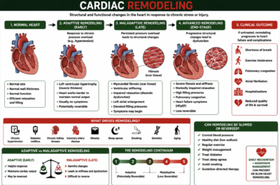
Cardiac remodeling is the heart’s response to chronic injury or long-term stress, especially uncontrolled hypertension.
Initially, remodeling is actually helpful.
Your heart adapts by changing its muscle so it can continue pumping enough blood to meet your body’s needs. This allows cardiac output to be maintained despite increased workload.
Over time, however, this adaptation becomes harmful.
Instead of protecting the heart, remodeling becomes maladaptive, causing progressive damage that eventually leads to heart failure.
Think of it like carrying a heavy backpack every day.
At first your muscles become stronger.
But after years of carrying that extra weight, your back begins to hurt, your posture changes, and permanent damage develops.
The heart responds in much the same way.
Two Major Types of Cardiac Remodeling
1. Concentric Remodeling (Pressure Overload)
Concentric remodeling occurs when the heart must pump against high pressure.
The most common causes include:
– Long-standing hypertension
– Aortic stenosis
– Hypertrophic cardiomyopathy
What Happens?
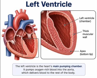
The left ventricular muscle becomes thicker.
While this sounds beneficial, there’s a problem.
As the muscle thickens:
The chamber actually becomes smaller
The heart becomes stiff
Filling during relaxation (diastole) becomes difficult
The pumping function (ejection fraction) may remain normal or even high, but the heart doesn’t fill with enough blood between beats.
This is called diastolic dysfunction and commonly leads to Heart Failure with Preserved Ejection Fraction (HFpEF).
Patients often experience:
– Shortness of breath
– Fatigue
– Exercise intolerance
– Swelling
Even though their EF may be 60–70%.
2. Eccentric Remodeling (Volume Overload)
Eccentric remodeling occurs when the heart is exposed to volume overload or muscle damage.
Common causes include:
– Dilated cardiomyopathy
– Previous heart attack
– Chronic mitral regurgitation
– Chronic aortic regurgitation
– Advanced HFrEF
What Happens?
Instead of becoming thicker…
The left ventricle begins to stretch and enlarge.
As the ventricle dilates:
Muscle fibers lengthen
The chamber becomes more spherical instead of elliptical
The heart becomes weaker
Stroke volume declines
Ejection fraction falls
Eventually, the heart simply cannot pump enough blood forward.
Why Remodeling Is Dangerous?
As remodeling progresses, the damage extends far beyond the heart muscle.
The ventricle enlarges and becomes progressively less efficient.
Wall Stress Increases
According to the Law of Laplace, a larger ventricle experiences greater wall stress.
More wall stress means:
Higher oxygen demand
Progressive weakening
Faster heart failure progression
The Mitral Valve May Begin to Leak
As the ventricle stretches, the mitral valve annulus also stretches.
This prevents the valve from closing completely, leading to functional mitral regurgitation, which places even more strain on the heart.
Scar Tissue Develops
Over time, damaged heart muscle is replaced with fibrosis (scar tissue).
Scar tissue:
– Doesn’t contract
– Doesn’t relax normally
– Cannot be reversed once extensive
Electrical Pathways Change
Fibrosis also disrupts the heart’s electrical system.
This increases the risk of:
– Ventricular tachycardia
– Ventricular fibrillation
– Sudden cardiac death
– Cardiac Output Eventually Falls
Although the body initially compensates, these mechanisms eventually fail.
The heart can no longer keep up with the body’s demands.
Can Remodeling Be Reversed?
Fortunately, yes—at least partially.
This is known as reverse remodeling.
With aggressive treatment, the heart can often become:
– Smaller
– Stronger
– More efficient
https://www.buzzsprout.com/765992/episodes/19517476
Reverse remodeling has been demonstrated with guideline-directed medical therapy.
These therapies include: 4 pillars of medications that are taken together and optimized, CRT, etc.
1. Sacubitril/valsartan (Entresto) or ACE inhibitors/ARBs if you have angioedema or entresto is not tolerated
2. Evidence-based beta blockers: Carvedilol or Metoprolol succinate or Bisoprolol
3. Mineralocorticoid receptor antagonists: Spironolactone or Eplerenone
5. SGLT2 inhibitors: Empagliflozin or Dapagliflozin
Cardiac resynchronization therapy (CRT) in appropriate patients
Correction of reversible causes such as:
– Severe valve disease
– Tachycardia-mediated cardiomyopathy
– Uncontrolled hypertension
Patients who respond well often demonstrate:
– Smaller left ventricular dimensions (decreased LVIDd and LVIDs)
– Improved ejection fraction (for example, from 20% to 40–50%)
– Improved global longitudinal strain (GLS)
– Reduced left atrial enlargement
– Better exercise tolerance
– Fewer heart failure symptoms
Why Blood Pressure Matters So Much?
Every heartbeat pushes blood against the arterial walls.
The higher the blood pressure…
The harder the heart must work.
Day after day.
Year after year.
Even modest elevations in blood pressure accelerate remodeling.
The good news is that lowering blood pressure reduces this stress before permanent damage occurs.
The 2017 ACC/AHA Blood Pressure Targets
The 2017 American College of Cardiology (ACC)/American Heart Association (AHA) Guideline for the Prevention, Detection, Evaluation, and Management of High Blood Pressure in Adults recommends individualized blood pressure goals based on cardiovascular risk.
Blood Pressure Goal: Less Than 130/80 mm Hg
A target of <130/80 mm Hg is recommended for most adults with hypertension, particularly those with: Established cardiovascular disease (coronary artery disease, stroke, peripheral artery disease) Heart failure Chronic kidney disease Diabetes mellitus Adults with hypertension who have a 10-year ASCVD risk ≥10% Most adults being treated for hypertension to reduce cardiovascular events Maintaining blood pressure below 130/80 mm Hg reduces the risk of:
Heart attack
Stroke
Heart failure
Kidney disease progression
Cardiovascular death
When Is a Goal Closer to Less Than 120/80 mm Hg Appropriate?
Although the 2017 ACC/AHA guideline generally recommends <130/80 mm Hg as the treatment goal, evidence from the SPRINT trial supports a more intensive systolic blood pressure target of <120 mm Hg in carefully selected, high-risk adults when it can be achieved safely. This approach may be appropriate for some patients, including:
– Adults at high cardiovascular risk without diabetes
– Individuals with chronic kidney disease (in selected cases)
– Older adults (55 or older) who tolerate intensive treatment without significant side effects
– Patients managed by clinicians who can monitor for low blood pressure, kidney function changes, and electrolyte abnormalities
An intensive target is not appropriate for everyone. It requires individualized decision-making, as more aggressive treatment may increase the risk of dizziness, falls, hypotension, or kidney-related side effects in some patients.
What Does Remodeling Look Like on an Echocardiogram?
Advanced heart failure specialists often monitor remodeling using measurements such as:
– LVIDd (Left Ventricular Internal Diameter in Diastole): An enlarged measurement suggests ventricular dilation.
– LVIDs (Left Ventricular Internal Diameter in Systole): Helps assess pumping function.
– Global Longitudinal Strain (GLS): Detects impaired heart muscle contraction, often before the ejection fraction declines.
– Left Atrial Size: Enlargement suggests chronically elevated filling pressures and long-standing diastolic dysfunction.
– Ejection Fraction (EF): Measures how much blood the left ventricle pumps out with each beat.
For example, a patient with:
– LVIDd of 6.72 cm
– Severely abnormal GLS
– Enlarged left atrium
– Reduced EF
shows a pattern consistent with advanced eccentric remodeling, commonly seen in dilated cardiomyopathy or advanced heart failure with reduced ejection fraction (HFrEF). These measurements help clinicians assess disease severity and monitor the response to guideline-directed medical therapy over time.
The Bottom Line
– High blood pressure doesn’t just raise a number on a cuff—it slowly changes the structure and function of your heart.
– By maintaining your blood pressure at less than 130/80 mm Hg, and in selected high-risk individuals aiming for a systolic pressure closer to 120 mm Hg under medical supervision, you can reduce stress on your heart, slow or prevent harmful remodeling, and significantly lower your risk of heart attack, stroke, heart failure, kidney disease, and premature death.
– The best time to protect your heart is before remodeling becomes permanent. Controlling your blood pressure today is one of the most effective investments you can make in your long-term cardiovascular health.
References
1. American College of Cardiology Foundation, American Heart Association, & Heart Failure Society of America. (2022). 2022 AHA/ACC/HFSA guideline for the management of heart failure. Circulation, 145(18), e895–e1032. https://doi.org/10.1161/CIR.0000000000001063
Bozkurt, B., Colvin, M., Cook, J., Cooper, L. T., Deswal, A., Fonarow, G. C., Francis, G. S., Lenihan, D., Lewis, E. F., McNamara, D. M., Pahl, E., Vasan, R. S., & Reddy, V. (2016). Current diagnostic and treatment strategies for specific dilated cardiomyopathies. Circulation, 134(23), e579–e646. https://doi.org/10.1161/CIR.0000000000000455
2. Lang, R. M., Badano, L. P., Mor-Avi, V., Afilalo, J., Armstrong, A., Ernande, L., Flachskampf, F. A., Foster, E., Goldstein, S. A., Kuznetsova, T., Lancellotti, P., Muraru, D., Picard, M. H., Rietzschel, E. R., Rudski, L., Spencer, K. T., Tsang, W., & Voigt, J. U. (2015). Recommendations for cardiac chamber quantification by echocardiography in adults: An update from the American Society of Echocardiography and the European Association of Cardiovascular Imaging. Journal of the American Society of Echocardiography, 28(1), 1–39.e14. https://doi.org/10.1016/j.echo.2014.10.003
3. McDonagh, T. A., Metra, M., Adamo, M., Gardner, R. S., Baumbach, A., Böhm, M., Burri, H., Butler, J., Čelutkienė, J., Chioncel, O., Cleland, J. G. F., Coats, A. J. S., Crespo-Leiro, M. G., Farmakis, D., Gilard, M., Heymans, S., Hoes, A. W., Jaarsma, T., Jankowska, E. A., et al. (2021). 2021 ESC Guidelines for the diagnosis and treatment of acute and chronic heart failure. European Heart Journal, 42(36), 3599–3726 (tel:3599-3726). https://doi.org/10.1093/eurheartj/ehab368
4. Nagueh, S. F., Smiseth, O. A., Appleton, C. P., Byrd, B. F., Dokainish, H., Edvardsen, T., Flachskampf, F. A., Gillebert, T. C., Klein, A. L., Lancellotti, P., Marino, P., Oh, J. K., Popescu, B. A., & Waggoner, A. D. (2016). Recommendations for the evaluation of left ventricular diastolic function by echocardiography. Journal of the American Society of Echocardiography, 29(4), 277–314. https://doi.org/10.1016/j.echo.2016.01.011
Otto, C. M. (2023). The practice of clinical echocardiography (6th ed.). Elsevier.
5. Ponikowski, P., Voors, A. A., Anker, S. D., Bueno, H., Cleland, J. G. F., Coats, A. J. S., Falk, V., González-Juanatey, J. R., Harjola, V. P., Jankowska, E. A., Jessup, M., Linde, C., Nihoyannopoulos, P., Parissis, J. T., Pieske, B., Riley, J. P., Rosano, G. M. C., Ruilope, L. M., Ruschitzka, F., et al. (2016). 2016 ESC Guidelines for the diagnosis and treatment of acute and chronic heart failure. European Heart Journal, 37(27), 2129–2200 (tel:2129-2200). https://doi.org/10.1093/eurheartj/ehw128
6. Towbin, J. A., McKenna, W. J., Abrams, D. J., Ackerman, M. J., Calkins, H., Darrieux, F. C. C., Daubert, J. P., de Chillou, C., Elliott, P., Hamilton, R. M., Hershberger, R. E., Judge, D. P., Leong, K. M. W., Mestroni, L., Priori, S. G., et al. (2019). 2019 HRS expert consensus statement on evaluation, risk stratification, and management of arrhythmogenic cardiomyopathy. Heart Rhythm, 16(11), e301–e372. https://doi.org/10.1016/j.hrthm.2019.05.007
7. Voigt, J. U., Pedrizzetti, G., Lysyansky, P., Marwick, T. H., Houle, H., Baumann, R., Pedri, S., Ito, Y., Abe, Y., Metz, S., Song, J. H., Hamilton, J., Sengupta, P. P., Kolias, T. J., d’Hooge, J., Aurigemma, G. P., Thomas, J. D., & Badano, L. P. (2015). Definitions for a common standard for two-dimensional speckle-tracking echocardiography. Journal of the American Society of Echocardiography, 28(2), 183–193. https://doi.org/10.1016/j.echo.2014.11.003
8. Yancy, C. W., Jessup, M., Bozkurt, B., Butler, J., Casey, D. E., Colvin, M. M., Drazner, M. H., Filippatos, G. S., Fonarow, G. C., Givertz, M. M., Hollenberg, S. M., Lindenfeld, J., Masoudi, F. A., McBride, P. E., Peterson, P. N., Stevenson, L. W., & Westlake, C. (2017). 2017 ACC/AHA/HFSA focused update of the 2013 ACCF/AHA guideline for the management of heart failure. Circulation, 136(6), e137–e161. https://doi.org/10.1161/CIR.0000000000000509
Braunwald, E. (2022). Braunwald’s heart disease: A textbook of cardiovascular medicine (12th ed.). Elsevier.
9. Zipes, D. P., Libby, P., Bonow, R. O., Mann, D. L., & Tomaselli, G. F. (Eds.). (2022). Braunwald’s heart disease: A textbook of cardiovascular medicine (12th ed.). Elsevier.